Anatomy & Physiology
External anatomy:
All Sipunculids are unsegmented, and maintain a vermiform bauplan. This body plan consists of two main sections: introvert & trunk. At the tip of the introvert is the tentacular crown, and the mouth. P. nigrescens has a rudimentary tentacular crown consisting of only nuchal tentaces. These are limited to a dorsal arc surrounding the nuchal organ (Cutler, 1994). All members of the Phascolosoma genera maintain the presence of laterally compressed angled hooks arranged in rings around the base of the introvert. Although the introvert hooks of P. nigrescens are often key in indentifying it from members of different species, high variability exists in the morphology of these (Cutler, 1990). Figure 12 represents a small subset of variability in introvert hooks of P. nigrescens. The full degree of morphological variability extends much further than the seven examples provided (Cutler, 1990). These hooks are often present in a series of rings. In P. nigrecens, they usually exist in a series of over 50 incomplete rings at the base of the introvert (Edmonds, 1955; Cutler, 1994).
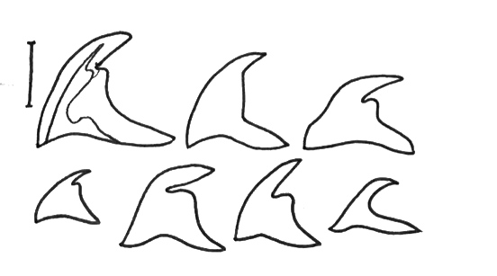 Figure 12: Subset of variability in introvert hook shapes of P. nigrescens subset includes collections from populations in Hawai'i, Madagascar, & Japan. All variations maintain the same internal structure, only shown in the first hook drawing. Scale line = 50um, adapted from Cutler 1990
The dorsal side of P. nigrescens’ introvert is often striped with black or brown patches with varying sizes. In all other species, the presence of these pigment bands is consistent, except for P. nigrescens (Cutler, 1990). P. nigrescens lack the presence of an anal shield. Present on the external epidermis of P. nigrescens are glandular structures known as papillae (Figure 14A). Three general shapes have been identified: domelike, mammillate, and conical. P. nigrescens has domelike papillae (Cutler, 1990). The function of these is not completely understood.
Internal anatomy:
Figure 13 depicts the internal anatomy of the Phascolosoma genera. P. nigrescens has a double-helix-shaped intestine spiral stretching from an oesophagus, located near the anterior end of the trunk, to the posterior end of the trunk, then ending at the anus. The anus is located at the anterior end of the trunk near the beginning of the introvert (Cutler, 1994). This odd placement may help ensure excrement does not fall inside the introvert when fully retracted. Directly below the anus is the rectal caecum, the purpose of which remains unknown (Cutler, 1994). The wing muscle serves to hold the terminal rectum on the body wall, and the spindle muscle extends from it towards gut coil. The spindle muscle serves to hold the intestinal coil in place, and ensure proper orientation. Sipunculids use a metanephridia to remove metabolic waste from the body. These are present in the trunk of P. nigrescens, and open ventrolaterally at the anterior end of the trunk. Retraction of the introvert is controlled by four retractor muscles, which extend to the posterior end of the trunk. In most Phascolosoma two eyespots are present at the base of the tentacular crown (Saiz, 1993).
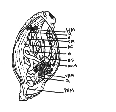 Figure 13: Internal view of typical Phascolosoma WM: wing muscle, R: rectum, N: nephridia, SM: spindle muscle, RC: rectal caecum, O: oesophagus, ES: eye spots, DRM: dorsal retractor muscle, VRM: ventral retractor muscle, G: gonads, PSM: posterior spindle muscle, adapted from Cutler, 1994
A frontal plane section of P. nigrescens from the New South Whales intertidal headlands was prepared, stained with phalloidin, and is depicted in Figure 14. Areas of interest have been labelled A - F. A & B depict the typical dome shaped epidermal papillae of P.nigrescens. C depicts both types of circular body wall muscles, and a section of a introvert retractor muscle (shown in D). E shows a section of the gut coil, and F depicts part of the metanephridia.
Figure 14: Frontal plane section of P. nigrescens stained with phalloidin. Images were captured on Olympus differential interference contrast (DIC; Nomarski) microscope. Composite image was created from 7 images taken with XScope using the PTGui free trial. Scale bar composite = 0.5cm; A=10um; B=100um; C=50um; D=100um; E=50um; F=100um.
P. nigrescens' central nervous system is made up of an anterior cerebral ganglion with a circumoesophageal connective and ventral nerve cord. The epithelium surrounding the cerebral ganglion contains bipolar sensory cells, so it is suggested it is not a larval vestige, but rather serves some sensory function (Purschke et al., 1997). The same study by Purschke (1997) suggest the nuchal organ, located at the apical tip of the introvert in P. nigrescens, is used for chemoreception. |